How does a digital impression work?
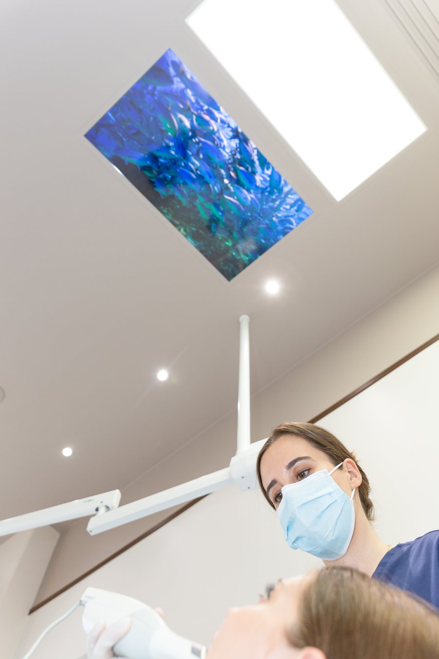
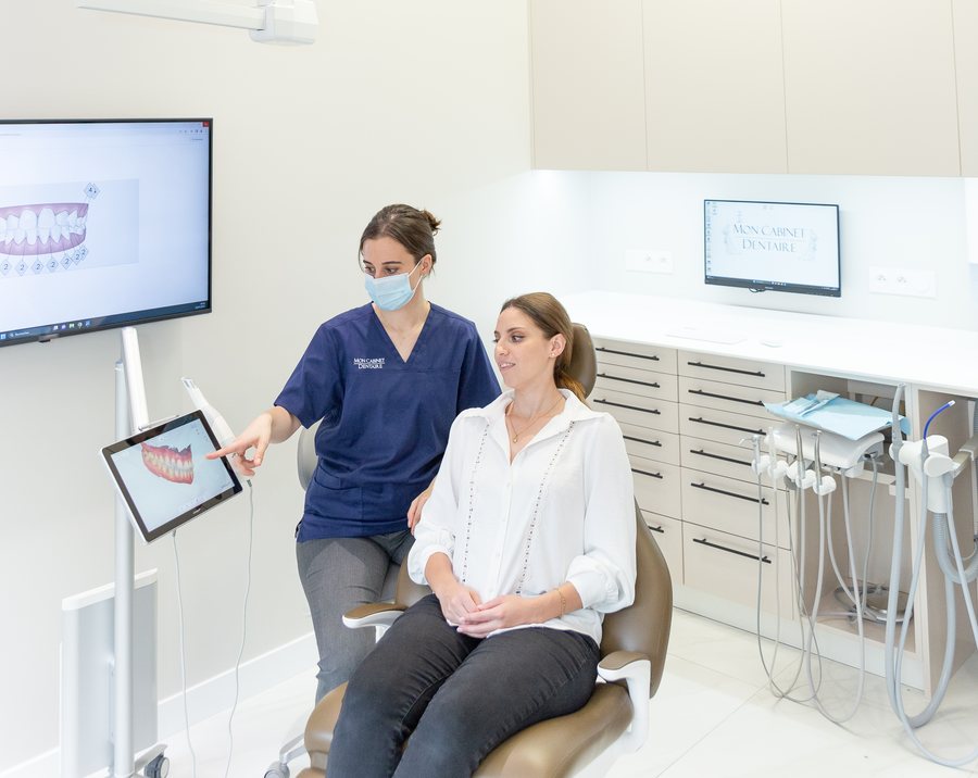
A small camera is moved along the teeth and gums. It records thousands of images per second and assembles them into a three-dimensional model, displayed in colour on the screen as it goes. The bite, that is the way your teeth meet, is recorded the same way.
The resulting model replaces the plaster cast: it is sent to the dental laboratory digitally, stored in your file and compared with later recordings if needed.
Why 3Shape
3Shape is a Danish company founded in 2000 in Copenhagen. Its TRIOS scanners and design software are used by a large number of practices and dental laboratories around the world; it is one of the reference manufacturers in the field, and the system we chose for the practice.
- True colour: the scan reproduces the shade of teeth and gums, with a colour measurement useful for ceramics.
- Software: the model is automatically cleaned of the soft tissues (tongue, cheeks) that get in the way.
- Open format: the file can be sent to any laboratory and any planning software.
- Built-in applications: simulation of an orthodontic treatment, monitoring over time by superimposing scans, smile design, design of splints and aligners.
What it changes for you
- Comfort: no more paste in the mouth or gag reflex; the recording can be paused and resumed.
- Accuracy: the digital model does not distort, unlike a silicone impression that is transported and then cast in plaster; crowns, veneers, onlays and retainers fit better.
- Understanding: you see your teeth in three dimensions on the screen, which makes explanations and decisions easier.
- Monitoring: by superimposing two scans, we measure wear, gum recession or tooth movement objectively.
- Exchanges: the laboratory receives the file immediately, with no transport or disinfection of a cast.
Which treatments do we use it for?
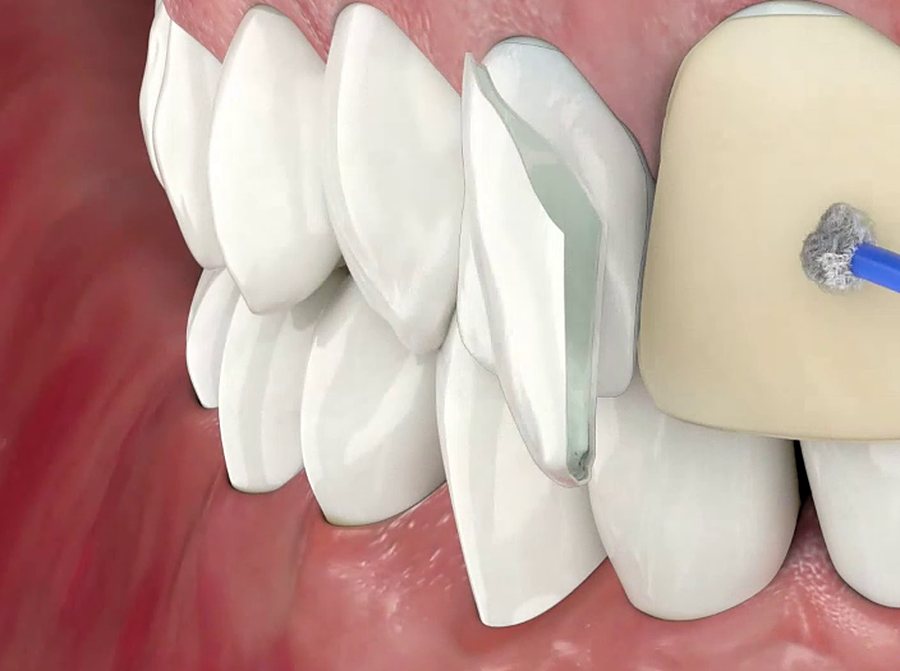
- Prosthetics: crowns, bridges, inlays and onlays, veneers, implant-supported restorations.
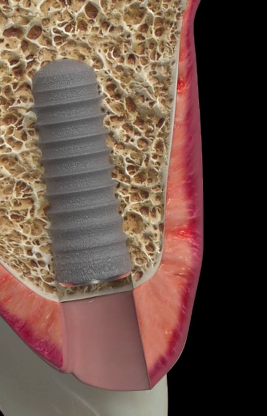
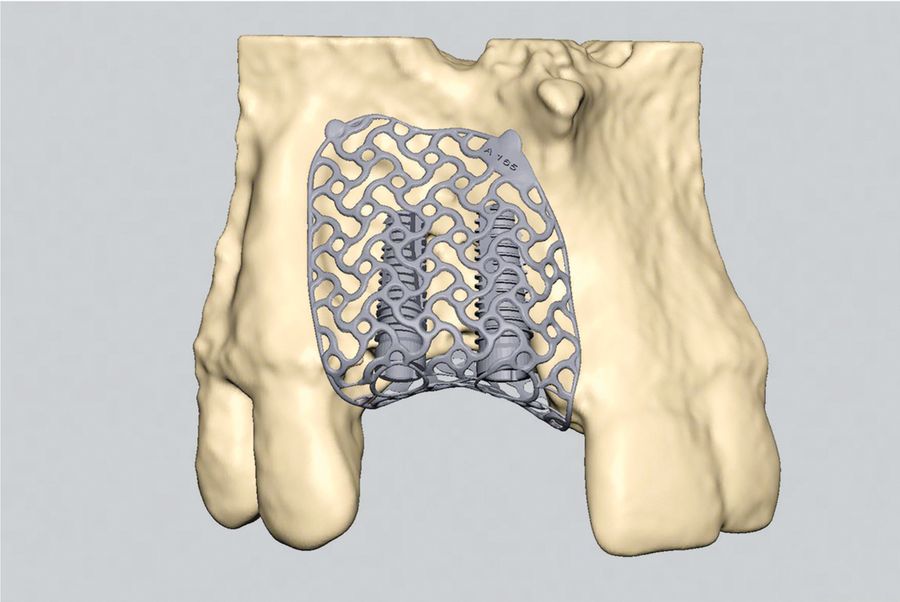
- Implantology: planning the position of implants and making surgical guides, by combining the optical model with three-dimensional imaging.
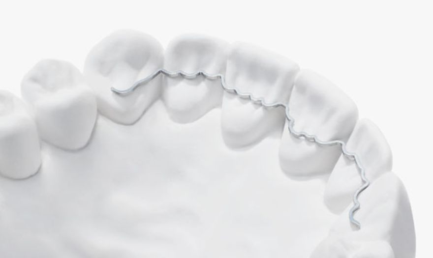
- Periodontics: custom-made retainers (Memotain wire), monitoring of gum recessions.
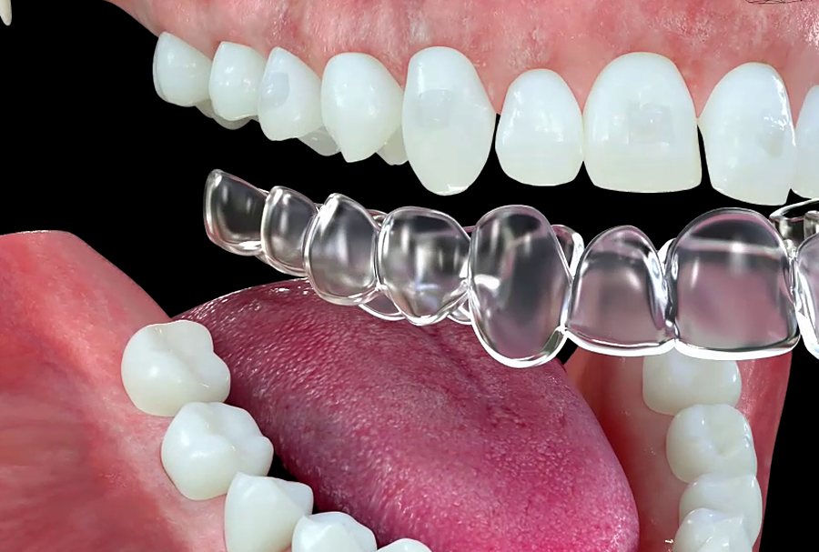
- Orthodontics: simulation of the result, clear aligners, retention at the end of treatment.
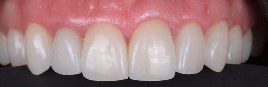
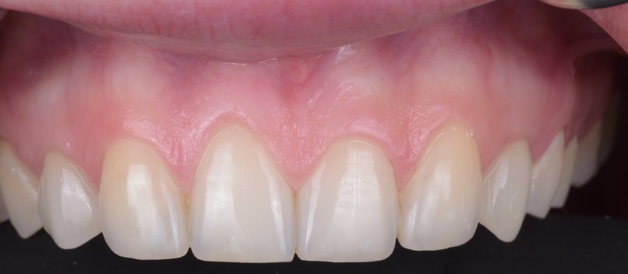
- Aesthetics: digital smile design and virtual try-in before veneers.
The limits
The scanner records what it sees: a preparation margin located deep under the gum or significant bleeding can hinder the reading, and a conventional impression remains possible. For some removable dentures, the traditional impression also keeps its place. The practitioner chooses the technique suited to each case.