
Specialty
Oral surgery
Oral and dental surgery: an essential pillar of oral and dental health.
Oral and dental surgery is a specialty of dentistry dedicated to the diagnosis and treatment of pathologies, abnormalities, and injuries affecting the mouth, teeth, jaws, and surrounding tissues. This specialty allows for the treatment of a wide variety of conditions, combining clinical expertise and advanced surgical techniques.
Dental extractions: a common surgical procedure
- Simple dental extractions
Simple extractions are performed when the teeth are visible and accessible in the mouth. They are often necessary in cases of:
- Deep caries: When the tooth is too damaged to be restored.
- Dental infection that cannot be treated.
- Advanced periodontal diseases: When the tooth is mobile due to bone loss.
- Orthodontic preparation: To create spaces in the dental arch for a realignment through orthodontic treatment.
This procedure is performed under local anesthesia and, thanks to modern tools, is quick and painless. Special attention is paid to preoperative instructions and post-operative follow-up to ensure proper healing and the least possible pain.
- Extractions of wisdom teeth
Wisdom teeth, also called third molars, are often a source of complications. When they are misaligned (incus or semi-incus), they can cause:
- Infections (pericoronaryites).
- Intense pain related to pressure on adjacent teeth.
- Cysts or bone lesions.
- Disalignment of the other teeth.
- Deep caries on the wisdom tooth or on adjacent teeth.
The extraction of wisdom teeth can be simple or complex, depending on their position in the alveolar bone and their proximity to sensitive structures, such as the inferior alveolar nerve or adjacent teeth. Complex extractions often require advanced surgical techniques and significant experience from the surgeon to minimize bone trauma and accelerate recovery.
Bone grafts: a crucial step for oral reconstruction
Bone loss in the jaw can occur as a result of periodontal diseases, old tooth extractions, or dental or bone trauma. Bone grafts allow for the restoration of the bone volume necessary to place dental implants in a durable and functional manner.
- Horizontal bone grafts
Horizontal grafts aim to widen the width of the alveolar bone in areas where it is too thin. This is often required when placing implants in the anterior region, where the thinness of the bone crest can compromise the stability of the implant. This technique uses materials such as autogenous bone (derived from the patient) or biomaterials.
- Vertical bone grafts
When the height of the bone is insufficient, particularly in the posterior areas of the jaw, a vertical graft is performed. This procedure is complex and requires meticulous planning on the part of the practitioner.
- Innovations in bone grafts
Modern techniques include the use of customized titanium grids and bioactive graft materials that promote rapid regeneration and better integration of the grafted bone. These innovations guarantee optimal aesthetic and functional results, even in the most complex cases.
4. Sinus grafts
Sinus fillings, also known as sinus elevations, are essential oral surgical procedures to prepare for the placement of dental implants in the posterior region of the upper jaw. When the bone height below the maxillary sinus is insufficient, these techniques allow for increased bone volume by adding graft material between the sinus floor and the sinus membrane.
Sinus reconstructions, and in particular the sinus lift technique via the lateral approach, represent a major advancement in oral surgery, enabling the rehabilitation of patients with severe bone loss. These procedures not only offer the possibility of placing dental implants in otherwise inaccessible areas, but also ensure long-term stability and function.
4.1 Sinus lift technique by lateral approach
Lateral sinus lift, or lateral sinus elevation, is a method particularly indicated in cases where the remaining bone height is very low, usually less than 5 mm.
Surgical procedure:
- incision and lateral access:
A small incision is made in the gum at the posterior region, followed by the creation of a lateral window in the bone wall of the maxillary sinus. This window allows direct access to the sinus membrane (Schneider membrane).
- Dissection of the sinus membrane:
Using specific instruments, the sinus membrane is gently lifted and separated from the bone floor, creating an area to accommodate the graft material.
- Introduction of the graft material:
An osseous material, often composed of autogenous bone, allogenic bone, or synthetic substitutes, is introduced into this newly created space. This graft will promote natural bone regeneration over time.
- Closing the window:
The bone window can be covered with a resorptive membrane to protect the grafted area, before suturing the gum to ensure good healing.
Specific indications for the sinus lift by the lateral approach
- When severe bone resorption makes the use of the crestal technique (Summers technique) impossible.
- In cases where a significant increase in bone volume is required (several millimeters).
- When the sinus membrane is thick enough and intact to allow for a safe lifting.
Advantages of the lateral approach technique
- Significant increase in bone volume: This technique allows for sufficient bone height to be achieved, even in cases where the residual bone is extremely limited.
- Optimum preparation for implants: Thanks to the addition of graft material, it guarantees a solid and durable base for dental implants.
- Possibility of combining with other interventions: In some cases, this technique can be performed simultaneously with the placement of implants, thus shortening the overall treatment time.
- Sustainability of results: The new bone formed provides long-term stability for the implants, reducing the risk of failure.
4.2 Sinus elevation by the crestal route
In comparison, the crestal technique (Summers technique) is indicated when the residual bone height is moderate (greater than 5 mm). It is performed via an access from the implant site, without a lateral opening. This method is less invasive, but is not suitable for cases of significant bone resorption.
It allows, during a minimally invasive procedure, to raise the sinus membrane by a few millimeters with the simultaneous placement of an implant.
5. Bone grafts by guided bone regeneration.
Guided bone regeneration (ROG) is an advanced technique in dental surgery aimed at restoring bone volume in areas where it is insufficient, particularly in preparation for the placement of dental implants. This method relies on the use of barrier membranes that promote selective bone growth while preventing the invasion of soft tissues.
Guided bone regeneration is an effective technique for restoring bone volume in specific situations, offering a less invasive alternative to traditional bone grafts. In-depth clinical evaluation is essential to determine the most appropriate method based on the individual needs of each patient.
Principles of guided bone regeneration:
- Use of barrier membranes: These membranes, whether resorptive or non-resorptive, are placed at the site of the graft to isolate the area of bone while the surrounding tissues are regenerating. This allows the bone cells to colonize the space and form a new bone without interference from the soft tissues.
- Application of graft materials: Bone substitutes, whether autogenous (derived from the patient), allogenic, xenogenic or synthetic, are used to fill in the bone defect. These materials serve as a support for the formation of new bone by stimulating the natural regeneration processes.
Indications from ROG:
- Localized bone defects: ROG is particularly indicated for small to medium bone losses, such as periapical defects or limited bone resorption.
- Preparation for implantation: When the bone volume is insufficient for the placement of an implant, ROG allows for the reconstruction of the bone necessary to ensure the stability and durability of the implant.
Advantages of ROG:
- Minimization of interventions: Unlike bone grafts in a single operation, the ROG is less invasive and reduces the risks associated with bone harvesting.
- Predictable results: With proper planning and controlled technique, the ROG offers high success rates and satisfactory osseous integration.
Limits of ROG:
- Limited regeneration volume: The ROG is more suitable for small to medium-sized bone defects. For larger reconstructions, more complex grafting techniques may be necessary.
- Healing time: Bone regeneration requires a healing time that can range from several months before the implant is placed.
Endodontic surgery: a surgical procedure to preserve your teeth.
Endodontic surgery is a specialized procedure aimed at treating infections or lesions located at the end of the tooth roots, especially when conventional endodontic treatments (such as root canal treatments) are not sufficient to resolve the problem. This approach allows the natural tooth to be preserved by eliminating persistent infectious foci.
Main interventions in endodontic surgery:
- Apicectomy: This procedure involves removing the end of the affected root and the surrounding infected tissues. After the ablation, a filling material is placed to seal the root canal hermetically, thus preventing any further infection.
- Radical resection: Primarily used for teeth with multiple roots, this technique involves removing a damaged or infected root while preserving the rest of the functional tooth.
- Apical curettage: This procedure aims to clean and disinfect the area around the apex of the root, removing infected tissue without necessarily removing part of the root itself.
Indications for endodontic surgery:
- Failure of conventional endodontic treatment: When the symptoms persist despite a properly performed root canal treatment.
- Complex radicular anatomy: Presence of calcified, curved, or inaccessible radicular canals by traditional methods.
- Presence of persistent apical lesions: cysts or granulomas that do not resolve after a conventional endodontic treatment.
Advantages of endodontic surgery:
- Preservation of the natural tooth: It prevents extraction and the use of prostheses or implants.
- Targeted infection elimination: Direct access to the infected area for effective disinfection.
- Quick recovery: With modern techniques, healing is usually quick and complications are rare.
It is essential to consult a dentist who specializes in endodontics to assess the need for endodontic surgery and determine the most appropriate procedure for each clinical situation.
Orthodontic surgery: An essential support for dental alignment
Orthodontic surgery plays a crucial role in the preparation or accompaniment of orthodontic treatments. These interventions allow for the resolution of anatomical obstacles or the optimization of the dental movements necessary for optimal alignment. Orthodontic surgery, whether involving the traction of incisors, frenectomy, or the placement of mini-vis, optimizes the results of orthodontic treatments by overcoming anatomical or mechanical obstacles. These interventions, carried out in close collaboration between the dentist and orthodontist, ensure a comprehensive and personalized approach for each patient.
Here is an overview of the main procedures:
- The inclusion of canine traction
The incisors, that is, those that remain stuck in the bone or the gum instead of emerging normally, are a common cause of dental misalignment. The surgery aims to facilitate their placement in the dental arch.
Procedure:
- A small incision is made to expose the included canine.
- An orthodontic device, such as an elastic chain or a button, is attached to the tooth to allow for its progressive traction under the supervision of the orthodontist.
Advantages:
- It allows the natural tooth to be preserved in the jawbone.
- It prevents complications such as resorption of the roots of nearby teeth or follicular cyst.
Indications:
- Inclusion of the canine detected radiologically.
- Absence of natural movement despite sufficient space in the arch.
- The frenectomy
Franektoomy is a surgical procedure consisting of removing or modifying a buccal frenum (tongue or lip) that interferes with oral function or aesthetics.
Orthodontic indications:
- Thick upper labial frenulum: It can cause a diastema (spacing between the central incisors) or prevent its closure.
- Restricted lingual frenulum (ankyloglossia): It can restrict tongue movements, affect speech, or interfere with certain orthodontic treatments.
Procedure:
- The vein is cut with local anesthesia using a surgical laser to minimize bleeding and accelerate healing.
- Post-operative rehabilitation is quick, with simple hygiene instructions.
- The placement of mini-orthodontic appliances
Minivisor orthodontics, or temporary brackets, are innovative devices used to provide stable support while the teeth are being moved. These small titanium screws, similar to small implants, are inserted into the maxillary or mandibular bone.
Indications:
- Necessity of complex dental movements (intrusion, extrusion, regression or advancement).
- Cases where neighboring teeth do not allow for sufficient anchorage.
- Reduction of parasitic forces that could affect other teeth.
Procedure:
- The minivis are inserted under local anesthesia in specific areas, depending on the necessary movements.
- Once the orthodontic treatment is complete, the braces are easily removed.
Advantages:
- They provide a fixed and precise anchorage, independent of the adjacent teeth.
- Reduce the need for bulky equipment.
- Certain types of orthodontic movements accelerate.
Conclusion
Oral and dental surgery plays a fundamental role in the management of oral pathologies and the reconstruction of the oral structures. Whether for extractions, implants, or bone grafts, each intervention is performed with expertise, taking into account the specific needs of each patient and relying on the most advanced technologies.
Your specialist
Dr Victor Hazout
Periodontist, Dentist
Dr. Michael Arroche
Dentist
Dr. Charles Anton
Dentist
OSSEOUS GRAFFIT PRE-IMPLANTATION
THE COMPLEX TEETH OF WISDOM
OSSEOUS COMPRESSION SINUSITIS
When the teeth are absent, the sinus swells, takes up more space, and there is no longer enough bone height to place an implant. Therefore, a sinus filling must be performed.
Sinus augmentation involves grafting a bone substitute into the lower base of the maxillary sinus in order to restore sufficient bone height for the placement of dental implants in a person with missing teeth in the posterior areas of the upper jaw.
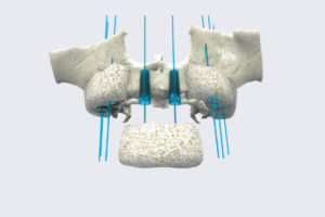
INNOVATION: 3D DESIGNATION OF THE BONE CRANIUM
This technique allows:
– A 3D design upstream of the transplant
– An optimal adaptation of the donor graft on the recipient bone
– A surgical procedure with a reduced duration of the intervention
– An improvement in patient comfort with less post-operative pain
My Dental Clinic
My Dental Clinic
119 Rue du Président Wilson
Ground floor on the right
92300 Levallois-Perret
contact@mon-cabinetdentaire.com
Tél: 01 42 67 05 04
Monday-Friday: 9:00 a.m. to 7:00 p.m.
Sam: 9:00 a.m. to 4:00 p.m.